
Back in December 2020 I wrote about a strange little instrument I had acquired from AllBids. It was a Cambridge Thermionic Corporation Model B Quartz Monochromator. There had been some discussion about it on a mailing list and, being sufficiently unusual, I just had to have it.
I played with it, took some photos, worked out roughly what it did and wrote a short article about it. Then I put it on a shelf and largely forgot about it.
A couple of months later I received an email from Mark Hugo in the USA. Mark had been looking for one of these instruments and had found my obscure little page while searching the Internet. Mine was sitting on a shelf doing nothing. Mark actually had a use for it, so I sent it to him.
How does it work?
I originally thought the monochromator worked using a diffraction grating. It doesn’t. Instead, it uses a rather clever property of quartz. When polarised light passes through quartz, its polarisation is rotated. Importantly, different wavelengths are rotated by different amounts.
The Model B uses several carefully cut pieces of quartz together with polarising filters. Turning the wavelength dial rotates parts of this optical system and selects a narrow band of light to pass through. The Model B has a bandwidth of about 15 nm. So rather than spreading white light into a rainbow and selecting one part of it, this little box uses quartz and polarisation to select the light it wants.
Not long after I sent it to Mark, photos started arriving.
One showed his microscope setup on a workbench. Sitting beside the microscope was a familiar black box. There was my monochromator, now on the other side of the world and actually doing something useful.
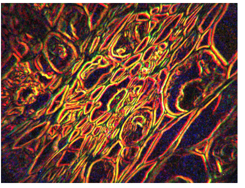
Mark was experimenting with monochromatic illumination and high-resolution microscopy. He was particularly interested in whether combining images taken at slightly different focal points could reveal detail that was difficult to see in a normal microscope image.
One set of images used 555 nm light, in the green part of the spectrum, at 1000× optical magnification.
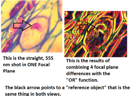
Mark then took images at different focal planes and processed the differences between them. In one example he combined four of these difference images using an OR operation.
The result was quite striking. Fine structures appeared which were barely visible in the original image.
Mark was also sensibly suspicious of what he was seeing. He wrote:
“I’ve been afraid I might be getting some ‘artifact’ causing these ‘fine lines’.”
That is an important question. Image processing can reveal information hidden in an image, but it can also create features which look convincing but are not really there.
Mark continued refining his process. He estimated some of the fine structures at around 60 to 70 nm wide and described his results as a possible “beyond Abbe” improvement.
Beyond Abbe
There is a physical limit to how much detail a conventional optical microscope can resolve. It is known as the Abbe diffraction limit and, with visible light, is generally around a couple of hundred nanometres depending on the microscope and wavelength being used.
That made Mark’s apparent 60 to 70 nm structures interesting. It does not prove that the microscope was resolving objects that small. A feature measuring 60 nm in a processed image is not necessarily the same thing as achieving 60 nm optical resolution. Mark clearly understood that and was actively trying to work out whether he was seeing real information or processing artefacts.
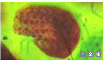
He also experimented with fluorescence under single-wavelength illumination. In one image using green light, he believed the strong red response was autofluorescence from the specimen.
Why I still write this stuff down
When I bought the monochromator, I had absolutely no use for it. It was simply an interesting piece of old scientific equipment. I wrote about it because that is largely why this website exists. I document strange equipment, things I build, things I repair and things I discover.
That little page then sat quietly on the Internet until someone on the other side of the world eventually searched for this fairly obscure instrument, found my page and contacted me. The monochromator went from sitting unused on my shelf to being part of an experimental microscope.
Before long Mark was sending me photos of the work he was doing with it. Who would have guessed. 🙂
And now, six years later, someone else has found that same old page while looking for information about one of these monochromators.
Which is probably as good a reason as any to start writing this stuff down again.
